Retinal detachment occurs when the retina separates from the underlying tissue, potentially causing permanent vision loss without prompt treatment. Symptoms include sudden flashes of light, new floaters, and shadows across the visual field, requiring immediate medical evaluation. Depending on the examination findings, retinal specialists may recommend pneumatic retinopexy, scleral buckling, or vitrectomy to repair the detachment. Here is more information about emergency procedures for retinal detachment:
Recognizing Retinal Detachment Symptoms
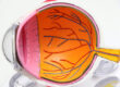
The retina lines the back of the eye and transmits visual information to the brain through the optic nerve. When the retina separates from underlying tissue, sudden vision changes may indicate a retinal detachment. Although detachment typically causes no pain, several symptoms require immediate medical evaluation. Common warning signs include:
- Noticing sudden floaters: Small specks, threads, or cobweb-like shapes appearing unexpectedly in the visual field.
- Seeing flashes of light: Brief flashes occurring in peripheral vision, particularly when the eye moves.
- Developing dark shadows: A curtain-like shadow spreading across part of the visual field.
- Experiencing blurred vision: Sudden changes in visual clarity or distortion affecting central or peripheral vision.
Patients experiencing these symptoms should seek immediate evaluation from an eye care provider or emergency department. Because detachment may progress without treatment, waiting for symptoms to disappear increases the risk of permanent vision loss. When contacting a provider, patients should describe when symptoms began, which eye is affected, and how vision has changed.
Understanding Treatment Options
Retinal detachment treatment involves procedures that reposition the retina and address underlying tears. Specialists select treatment methods based on the detachment’s location, severity, and underlying cause. Because retinal conditions vary, surgical approaches and recovery requirements differ between patients.
Pneumatic retinopexy involves injecting a gas bubble into the eye to help reposition the detached retina. Patients must maintain specific head positions while the bubble supports the retina during healing. Laser treatment or cryotherapy is also used to seal retinal tears and support reattachment. Alternatively, scleral buckling involves placing a silicone band around the eye to reduce traction on the retina.
Vitrectomy involves removing the vitreous gel and treating retinal tears or other abnormalities contributing to detachment. Surgeons may use a gas bubble or silicone oil to support the retina while healing occurs. Following surgery, patients receive instructions about positioning, activity restrictions, follow-up appointments, and recovery expectations.
Protecting Vision After Treatment
Recovery from retinal detachment surgery varies by procedure, and patients receive specific instructions regarding positioning, activity restrictions, and follow-up care. Patients with gas bubbles may need to maintain certain head positions and avoid air travel until their specialist approves. Follow-up examinations allow retinal specialists to monitor healing, evaluate retinal attachment, and identify possible complications. Because new floaters, flashing lights, or sudden vision changes may indicate complications, patients should seek immediate medical evaluation.
Schedule Your Appointment Today
Retinal detachment requires immediate medical evaluation because delayed treatment increases the risk of permanent vision loss. Sudden floaters, flashing lights, or dark shadows across the visual field may indicate a retinal tear or detachment. A dilated eye examination allows retinal specialists to assess the retina and determine appropriate treatment options. Contact a retinal specialist immediately for an urgent evaluation if you experience these warning signs.